Brief introduction to the lymphatic system:
The lymphatic system is a one-way drainage system, which works in close synergy with the cardiovascular system. Via an extensive network of lymphatic vessels, ducts and lymph nodes it recycles excess tissue fluid. This fluid accumulates between layers of tissue, because of normal capillary leakage. The walls of the lymph vessels act as one-way valves, only letting fluid (lymph) enter. Once in the vessels, the lymph is advanced towards the systemic circulation. Movement of lymph fluid depends on movement of muscles, contraction of the lymphatic vessels themselves and respiration. Along the way the lymph is filtered through several lymph nodes where it is inspected for foreign substances. This is an important part of the immune system, as here lymphocytes are produced, which help fight off invading bacteria. Upon reaching the thoracic duct the lymph re-enters the blood circulation.
Lymph: A clear fluid that contains proteins, fats, lymphocytes (white blood cells that fight infection and the growth of tumours), and plasma.
Lymph vessels: A network of thin walled tubes which help lymph flow through the body and return it to the bloodstream.
Lymph nodes: Small, bean-shaped structures that filter lymph and store white blood cells that help fight infection and disease. Lymph nodes are located along the network of lymph vessels found throughout the body. Clusters of lymph nodes are in the underarm, pelvis, neck, abdomen, and groin. Spleen, thymus, tonsils, and bone marrow are also part of the lymphatic system.
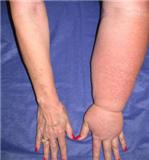 What is Lymphoedema?
A transport disorder of the lymphatic system: Lymphoedema (swelling) occurs when lymph flow is impaired. Either damage, obstruction or absence of vessels can lead to dysfunction of the lymphatic system. This can result in the accumulation and stagnation of lymph in the tissues. Most often limbs are affected, but lymph oedema can also occur in the face, neck, abdomen or genitals. Tissues with lymphoedema are at risk of infection and susceptible to progression. Lymphoedema is a treatable and manageable condition.
Types of Lymphoedema and Causes:
1. Developmental (inherited) disturbance of the lymphatic system (primary lymphoedema)
2. Acquired damage of lymphatic vessels and/or lymph nodes (secondary lymphoedema)
1. Primary Lymphoedema. Primary lymphoedema occurs in women more often than men. In most cases it is found on one side and is pronounced more distal. If it occurs on both sides, there is normally and asymmetry. Primary lymphoedema can be further classified according to the type of occurrence (congenital: already manifest at birth – lymphoedema praecox: occurring before the age of 35 – lymphoedema tardum: occurring after the age of 35)
2. Secondary Lymphoedema
Secondary lymphoedema can occur following surgical removal of lymph nodes or radiation therapy in the treatment of cancer. Secondary lymphoedema can also occur as a result of the following:
• Post-operative (e.g. after plastic or venous surgery).
• Post-traumatic (e.g. trauma which leads to injury of large lymph collectors such as open fractures, burns, wounds).
• Post-inflammatory (e.g. rheumatic diseases, sinusitis, recurrent phlebitis).
• Post-infection (e.g. recurrent cellulitis, inflammation of the lymph vessels, inflammation of lymph nodes).
Stages of Lymphoedema
Lymphedema progresses through stages, and treatment intervention in early stages (stage 0 and stage I) has been shown to result in very good treatment outcomes if managed appropriately
There are four stages of lymphoedema
Stage 0 (latent or subclinical )
In this stage the transport capacity of the lymphatic system is reduced, but the remaining lymph vessels are sufficient to manage the flow of lymph, and swelling is not visibly present.
Stage 1 (spontaneously reversible):
Tissue is still at the “pitting” stage, which means that when pressed by fingertips, the area indents and holds the indentation. Usually, upon waking in the morning, the limb(s) or affected area is normal or almost normal size.
Stage 2 (spontaneously irreversible):
The tissue now has a spongy consistency and is “non-pitting,” meaning that when pressed by fingertips, the tissue bounces back without any indentation forming). Fibrosis found in Stage 2 lymphedema marks the beginning of the hardening of the limbs and increasing size.
Stage 3 (lymphostatic elephantiasis):
At this stage the swelling is irreversible and usually the limb(s) is/are very large. The tissue is hard (fibrotic) and unresponsive; some patients consider undergoing reconstructive surgery called “debulking” at this stage.
When lymphedema remains untreated, protein-rich fluid continues to accumulate, leading to an increase of swelling and a hardening or fibrosis of the tissue. In this state, the swollen limb(s) becomes a perfect culture medium for bacteria and subsequent recurrent lymphangitis (infections). Moreover, untreated lymphedema can lead into a decrease or loss of functioning of the limb(s), skin breakdown, chronic infections and, sometimes, irreversible complications. In the most severe cases, untreated lymphedema can develop into a rare form of lymphatic cancer called Lymphangiosarcoma (most often in secondary lymphedema).
Treatment: According to the International Society of Lymphology, Combined Decongestive Therapy (CDT), also known as decongestive lymphatic therapy (DLT), is the treatment of choice for lymphoedema.
1. Manual Lymph Drainage – Patients receive Manual Lymph Drainage (MLD) to remove excess fluid and protein from the tissues. The MLD is performed to open lymphatics in the unaffected regions so these can help to drain the affected area. MLD stimulates lymphangions to increase their activity, which results in a decompression and emptying of obstructed lymphatic channels.
2. Compression Therapy – Multi-layered bandaging of the affected limb follows each MLD session. This is a precise and accurate procedure using specific bandages and interfacing materials.The bandages are applied exactly to conform to the patient’s tissues and are reapplied on a daily basis. They are short-stretch bandages that resist muscle contraction and are applied with comfortable padding underneath. The bandages help to maintain the reductions achieved with MLD and may even cause further reduction.
3. Exercise – Effective lymph flow depends on sufficient muscle and joint activity, especially if the functionality of the lymphatic system is compromised. Decongestive exercises are most effective if performed while the patient wears compression garments or bandages, which are also essential components in lymphedema management.
4. Skin Care and Hygiene – Good skin care plays an essential part in the treatment of lymphoedema. Daily skin cleansing with antibacterial washes and neutral balanced pH lotions will help to eliminate possible bacterial and fungal growth and so minimise the possibility of repeated attacks of cellulitis or lymphangitis.
5. Breathing – The lymph transport in the Thoracic Duct (Ductus Thoracicus) is mainly caused by the action of breathing. Taking into consideration that this duct transports 4 litres of lymph per day explains the importance of diaphragmatic breathing to help increase transport of lymphatic fluid.